Orders
Profile
How to Read Ultrasound Report of Pregnancy?

C-section & gynae problems · 4 years experience
AI-Generated Summary
Quick overview in 30 secondsSummary
- An ultrasound is a key part of prenatal care, used to assess fetal growth, monitor mother and baby's health, and detect any potential complications during pregnancy.
- There are five main types of ultrasound scans in pregnancy: transvaginal, standard, fetal echocardiography, 3-D ultrasound, and dynamic 3-D (4D) ultrasound, each serving different purposes.
- To read the report, observe the amniotic fluid (black area), baby (grey/white shape), and key measurements like CRL, head circumference, femur length, and abdominal circumference.
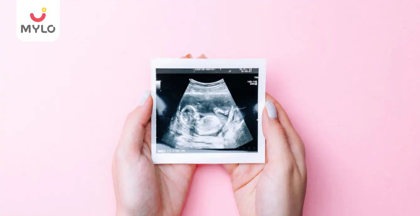
- Looking for trusted prenatal care essentials? Explore our Pregnancy Massage Oil + Coconut Oil - 200 ml each.


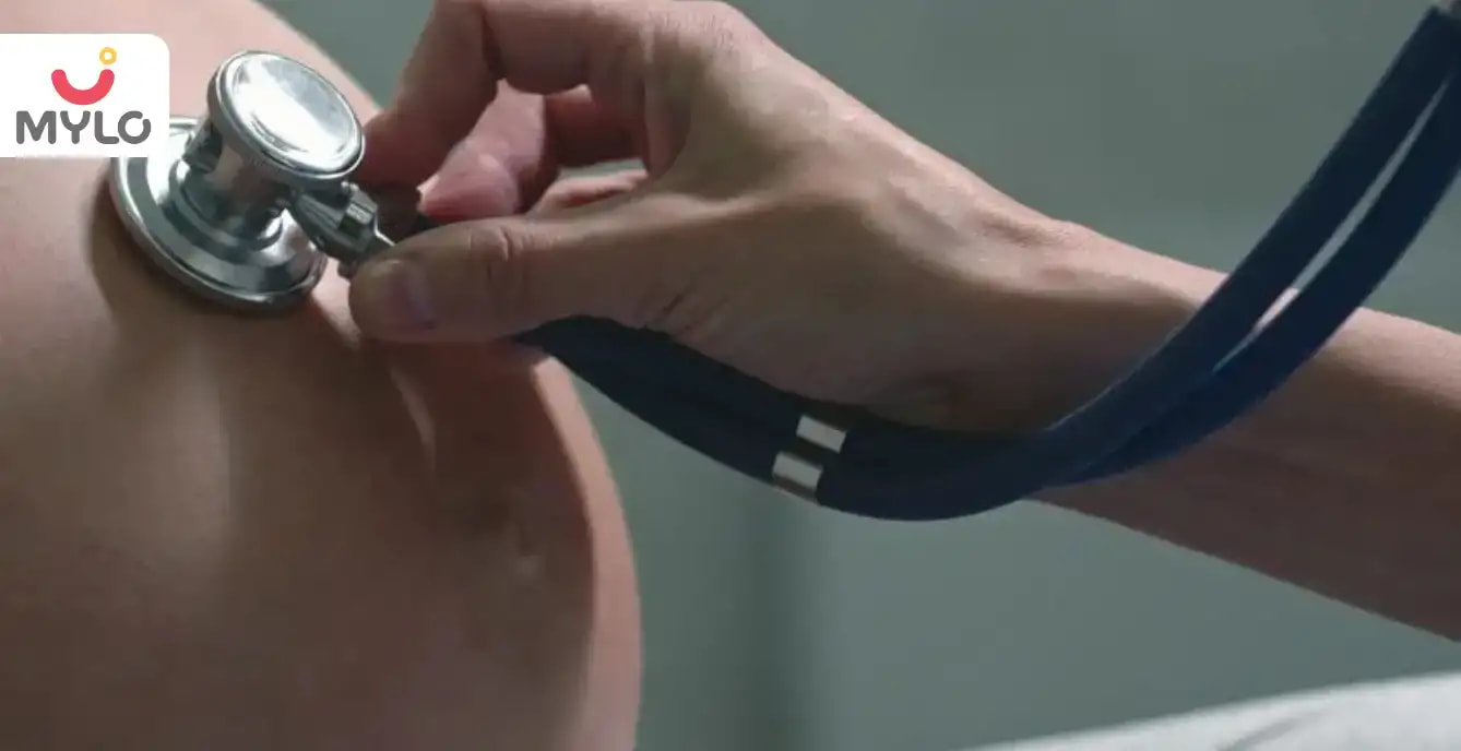
One of the things that excite and scare you the most during pregnancy is going for an ultrasound test. An ultrasound is a crucial component of prenatal care during pregnancy. Ultrasound scans are used to assess the growth and development of the fetus, monitor the health of the mother and baby, and detect any potential complications. However, an ultrasound report can be overwhelming and difficult to understand for many parents-to-be.
In this article, we will provide you with a comprehensive guide on how to read ultrasound report of pregnancy, including what to expect during the scan, the different types of ultrasound scans, and how to interpret the results. By the end of this article, you will have a clear understanding of what each section of the pregnancy ultrasound report means and how to discuss the findings with your healthcare provider.
What is an Ultrasound Scan?
Ultrasonography is a diagnostic procedure done during the three trimesters of pregnancy to assess the baby's growth, the health of the mother's reproductive organs, and the fetus's heart and confirm pregnancy status. An ultrasonography machine utilizes ultrasonic waves to visualize the inside of the womb through a specialized probe and ultrasound gel.
Types of Ultrasound in Pregnancy
With the advent of science and technology, ultrasonic scanning of the growing womb has made significant advancements. There are five major types of ultrasound in pregnancy:
1. Transvaginal ultrasound
‘Transvaginal’ means through the vagina. This scan is done for a mother-to-be who is facing a high-risk pregnancy or having any critical health concerns. It is generally done when the fetus is below 10 weeks.
2. Standard ultrasound
In this type of ultrasound, the technician moves a wand-shaped transducer over the stomach to see the 2D figure of your baby in the womb.
3. Fetal echocardiography
A fetal echo test is done to detect any issues or defects in the baby’s heart and if anything suspicious was detected with a standard ultrasound.
4. 3-D ultrasound
This sonography is done by a unique transducer that produces a complete and more detailed picture of your fetus.
5. Dynamic 3-D ultrasound (4D ultrasound)
This is similar to 3-D ultrasound. The difference here is that you can see the movement of your baby visually and can also have a video recorded in some cases.
How to Read Ultrasound Report of Pregnancy?
Using these steps, you will be able to comprehend your pregnancy ultrasound report and better understand your doctor:
1. Look at your womb
When you look at your womb in the sonogram, you will see a light gray or white line around the image's corners. Inside these lines, you will see a large black area known as amniotic fluid.
2. Look at your baby
Your baby will appear grey or white in the amniotic fluid, i.e., the dark area discussed above.
- In an ultrasound taken in the first 8 weeks of pregnancy, your fetus will be the size and shape of a baked bean.
- If you go for a scan at 12 weeks, you will see the head of the baby.
- At 20 weeks, the difference seen in the scan will be incredible. You will be able to locate the baby’s eyes, spine, heart, and feet.
Are the Numbers on the Ultrasound Picture Relevant to You?
The numbers written at the top of your scan report are data recorded by technicians that are relevant to themselves – your name, hospital reference codes, machine calibration settings, etc. When the technician starts taking the ultrasound of your uterus, the uppermost image of the scan will show the tissues above your uterus. You will also see the lining, the inner side, and the back part of your uterus in the ultrasound image.
Do Ultrasound Reading Colors Matter?
Yes, the reading colors matter. Usually, ultrasounds are black and white, but there is a subtle difference in the shade of each tone. The different tissues in your body transmit different sounds, some reflecting and other absorbing the waves. The density of tissues is directly proportional to how white they look. Hence, the variation in the shade of the scanned tissues is due to their varying densities. In the ultrasound of your uterus, your amniotic fluid will show up as a black area. Tissues will show up as grey. If ultrasounds were to be taken of solid tissues, such as bone, they would be seen as white.
You may also like: What Are the Common Tests You Will Have During Your Pregnancy?
Tips for Reading a Baby Sonogram
Here are tips for reading your baby's sonogram:
1. The gestational age of the fetus will be measured by CRL (crown-rump length). This is the measurement from the top to the bottom of baby's bum. Then, the resulting length is matched with an internal chart to determine the baby's gestational age inside the womb. Generally, this sonography is done between 7 weeks to 13 weeks of pregnancy.
2. Next in sonography, the head circumference is calculated. This calculation is done at 13 weeks of pregnancy.
3. Now, begins the measurement of the thighbone and femur bone. This is done to find out the longitudinal growth of the baby. This is also done at 13 weeks of pregnancy.
4. The fetus's height and length will be calculated by measuring the abdominal circumference. This is done at 20 weeks of pregnancy.
You may also like: What is BPD in Pregnancy?
The fetus measurements are rechecked to check for any abnormalities. A shorter than average humerus or the femur bone or missing fetal nasal bones are symptoms of Down Syndrome. This is done between 20–40 weeks of pregnancy.
Key Takeaways
In conclusion, reading an ultrasound report of pregnancy can be intimidating for many parents-to-be, but it is an essential part of prenatal care. Understanding the different types of ultrasound scans, what to expect during the procedure and how to read ultrasound report of pregnancy can help ease anxiety and ensure that you receive the best possible care for you and your baby. It is important to discuss any concerns or questions with your doctor as they can provide insight and guidance on the next steps in your pregnancy journey.
References
1. Ihnatsenka B, Boezaart AP. (2010). Ultrasound: Basic understanding and learning the language. Int J Shoulder Surg.
2. Ulrich CC, Dewald O. (2023). Pregnancy Ultrasound Evaluation. NCBI
Tags
How to Read Ultrasound Report of Pregnancy in Bengali, How to Read Ultrasound Report of Pregnancy in Telugu, How to Read Ultrasound Report of Pregnancy in Tamil
Popular Articles
Trending Articles
Loved by Mamas Navigating Their Pregnancy Journey
From scans to scares, the right prenatal care essentials make every milestone smoother and more reassuring for you and baby.
Pregnancy Massage Oil + Coconut Oil - 200 ml each


Baby Wellness Kit | Skincare Gift Set for Newborns


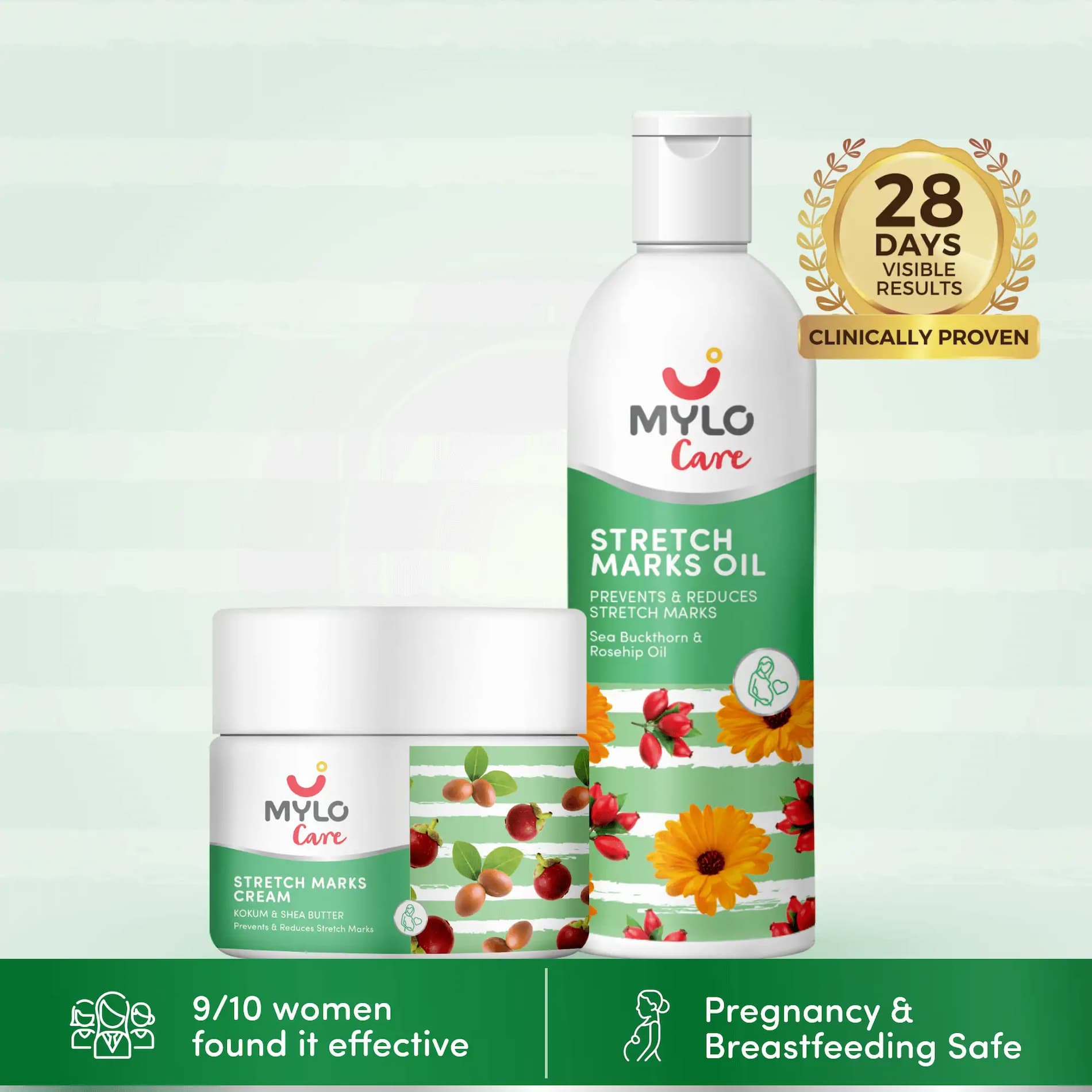
Stretch Marks Kit (Stretch Mark Oil + Stretch Marks Cream - 100ml each)
Made with Natural Ingredients | Tightens Skin | Prevents New and Fades Old Stretch Marks |


Article Posted Under
Related Articles


Related Topics
Medical Disclaimer
This content is for informational purposes only and should not replace professional medical advice. Consult with a physician or other health care professional if you have any concerns or questions about your health. If you rely on the information provided here, you do so solely at your own risk.


AWARDS AND RECOGNITION

Mylo wins Forbes D2C Disruptor award

Mylo wins The Economic Times Promising Brands 2022
AS SEEN IN


- Mylo Care: Effective and science-backed personal care and wellness solutions for a joyful you.
- Mylo Baby: Science-backed, gentle and effective personal care & hygiene range for your little one.
- Mylo Community: Trusted and empathetic community of 10mn+ parents and experts.
Product Categories
Baby Carrier | Baby Soap | Baby Wipes | Stretch Marks Cream | Baby Cream | Baby Shampoo | Baby Massage Oil | Baby Hair Oil | Stretch Marks Oil | Baby Body Wash | Baby Powder | Baby Lotion | Diaper Rash Cream | Newborn Diapers | Teether | Baby Kajal | Baby Diapers Pants | Cloth Diapers | Laundry Detergent | Lactation Granules |