Home

Scans & Tests

PCOS Ultrasound: What to Expect During the Procedure
In this Article

Scans & Tests
PCOS Ultrasound: What to Expect During the Procedure
Updated on 5 January 2024


Medically Reviewed by
Dr. Shruti Tanwar
C-section & gynae problems - MBBS| MS (OBS & Gynae)
View Profile

Polycystic Ovary Syndrome (PCOS) is a common hormonal disorder that affects many women worldwide. One of the diagnostic tools used to identify PCOS is a PCOS ultrasound. This non-invasive procedure allows doctors to visualize the ovaries and assess any abnormalities. If you're scheduled for an ultrasound, it's natural to have questions and concerns about what to expect during the procedure.
Who needs a PCOS ultrasound scan?
A PCOS ultrasound scan is typically recommended for women who exhibit symptoms of PCOS or have irregular menstrual cycles. Symptoms of PCOS may include irregular periods, excessive hair growth, acne, and difficulty getting pregnant.
If you're experiencing any of these symptoms or suspect you may have PCOS, your doctor may suggest an ultrasound to evaluate the condition of your ovaries and confirm the diagnosis.
You may also like : PCOS Acne: The Ultimate Guide to Causes, Treatment and Management
PCOS ultrasound vs normal ultrasound
While a normal ultrasound and a PCOS ultrasound may seem similar, there are some key differences between the two procedures.
A normal ultrasound is typically performed to examine various organs in the body, such as the liver, kidneys, or bladder. On the other hand, a PCOS ultrasound scan specifically focuses on the ovaries to identify any abnormalities or cysts.
During a PCOS ultrasound, the technician will carefully examine the size, shape, and appearance of the ovaries, as well as the presence of any cysts or follicles. This specialized ultrasound allows doctors to evaluate the condition of the ovaries and determine if PCOS is present.
The doctor may conduct a transvaginal ultrasound to diagnose PCOS. During a transvaginal ultrasound, a probe is inserted into the vagina to obtain detailed images of the ovaries and uterus. This procedure helps in visualizing the size and appearance of the ovaries, the presence of cysts, and the thickness of the endometrium.
You may also like: PCOS and Sex: Exploring Impact on Health and Debunking Common Myths
Is it necessary to do PCOS ultrasound during period?
It is not necessary to schedule a PCOS ultrasound during your period. In fact, it is often recommended to have the scan done a few days after your period has ended. This is because the menstrual cycle can affect the appearance of the ovaries, and performing the ultrasound during your period may make it more difficult to obtain accurate results.
Additionally, scheduling the ultrasound a few days after your period allows the technician to assess the ovaries when they are less likely to be affected by hormonal fluctuations. It's important to follow your doctor's instructions regarding when to schedule the ultrasound to ensure the most accurate results.
What is the best time to do ultrasound for PCOS?
The best time to schedule an ultrasound for PCOS is typically between days 5 and 10 of your menstrual cycle. This is when the ovaries are less likely to be affected by hormonal fluctuations and provide a clearer image for evaluation. However, it's important to consult with your doctor to determine the most appropriate timing for your specific situation.
They will take into account factors such as the regularity of your menstrual cycle and any additional symptoms you may be experiencing. By scheduling the ultrasound at the optimal time, you increase the chances of obtaining accurate results and a more comprehensive evaluation of your ovaries.
What to expect during a PCOS ultrasound?
Here’s what you can expect during the procedure:
-
During the ultrasound, you will be asked to change into a hospital gown and lie down on an examination table.
-
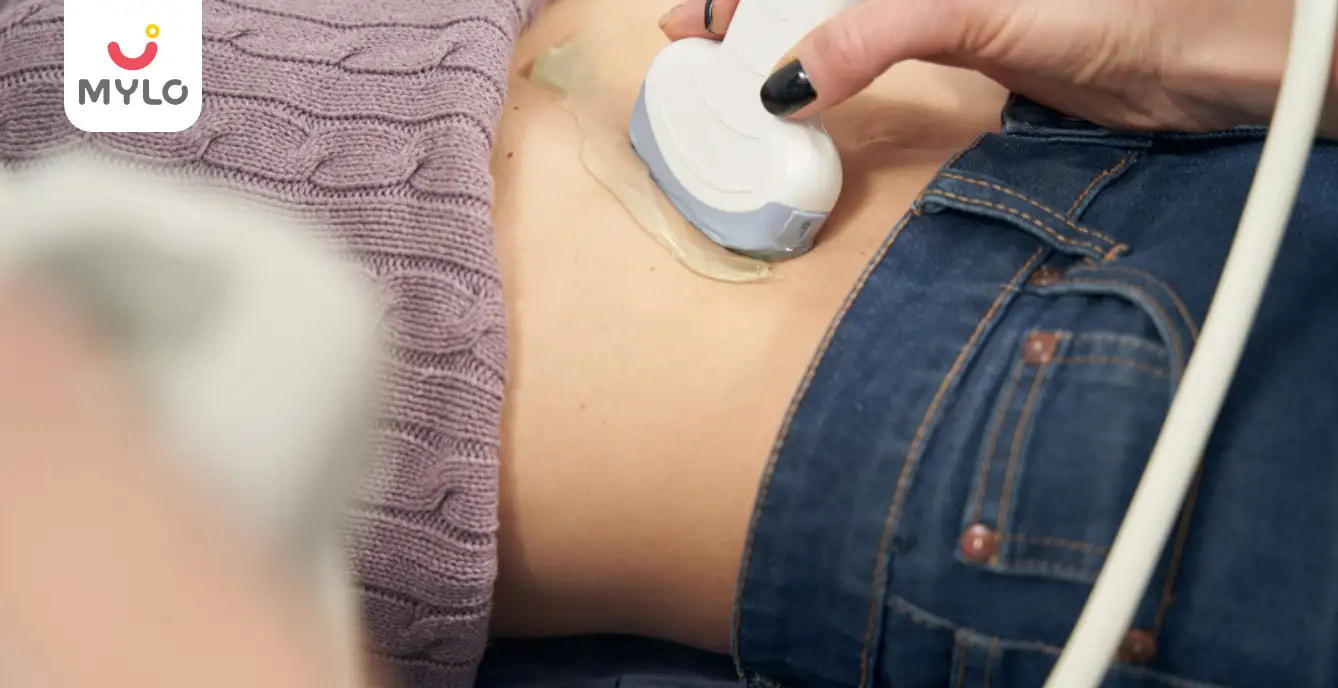
The technician will apply a gel to your abdomen and use a handheld device called a transducer to capture images of your ovaries.
-
The transducer emits sound waves that bounce off the structures in your body and create images on a screen.
-
The technician will move the transducer gently over your abdomen to obtain different views of the ovaries.
-
You may experience slight pressure or discomfort during the procedure, but it is generally well-tolerated.
-
The entire process usually takes around 30 minutes, after which you can resume your normal activities.
How to interpret a PCOS ultrasound report?
Interpreting a PCOS ultrasound report requires knowledge of the normal appearance of the ovaries and understanding the specific criteria used to diagnose PCOS. The report will typically include measurements of the ovaries, the number and size of any cysts or follicles, and the appearance of the ovarian tissue.
The presence of multiple small cysts and an increased ovarian volume are often indicative of PCOS. Your doctor will review the report, compare the findings to the diagnostic criteria, and provide you with an explanation of the results. It's important to consult with your doctor to fully understand the implications of the ultrasound report and discuss any necessary follow-up steps.
You may also like : Hysteroscopy: Everything You Need to Know About This Minimally Invasive Procedure
Tips to prepare for a PCOS ultrasound scan
Preparing for a PCOS ultrasound scan can help ensure a smooth and successful procedure. Here are five tips to help you prepare:
1. Follow your doctor's instructions
Your doctor will provide specific instructions on how to prepare for the ultrasound. It may include fasting for a certain period or drinking water before the scan. It's important to follow these instructions carefully to obtain accurate results.
2. Wear comfortable clothing
Choose loose-fitting and comfortable clothing to wear on the day of the scan. This will make it easier for the technician to access your abdomen during the procedure.
3. Stay hydrated
Drinking an adequate amount of water before the scan can help improve the clarity of the images obtained during the ultrasound.
4. Bring a support person
If you feel anxious or nervous about the procedure, consider bringing a support person with you. They can provide emotional support and help alleviate any concerns.
5. Ask questions
If you have any doubts or concerns about the procedure, don't hesitate to ask your doctor or the technician. They are there to ensure your comfort and address any questions you may have.
You may also like : Myomectomy: A Comprehensive Guide to Uterine Fibroid Removal Surgery
FAQs
1. Is PCOS obvious on ultrasound?
PCOS may not always be obvious on ultrasound. While the presence of multiple small cysts and an increased ovarian volume are common indicators of PCOS, other diagnostic criteria must also be considered. These include irregular menstrual cycles, elevated levels of androgens (male hormones), and clinical symptoms such as acne or excessive hair growth.
2. How is PCOS confirmed?
PCOS is typically confirmed through a combination of medical history, physical examination, blood tests, and imaging studies such as a PCOS ultrasound scan. Your doctor will evaluate your symptoms, perform necessary tests, and consider all the findings to arrive at a definitive diagnosis.
The Bottomline
A PCOS ultrasound is a valuable diagnostic tool in assessing the condition of the ovaries and confirming the presence of PCOS. Remember to consult with your doctor for personalized guidance and to address any concerns you may have.
References
1. Bachanek M, Abdalla N, Cendrowski K, Sawicki W. (2015). Value of ultrasonography in the diagnosis of polycystic ovary syndrome - literature review. J Ultrason.
2. Giménez-Peralta I, Lilue M, Mendoza N, Tesarik J, Mazheika M. (2022). Application of a new ultrasound criterion for the diagnosis of polycystic ovary syndrome. Front Endocrinol (Lausanne).


Medically Reviewed by
Dr. Shruti Tanwar
C-section & gynae problems - MBBS| MS (OBS & Gynae)
View Profile


Written by
Anupama Chadha
Anupama Chadha, born and raised in Delhi is a content writer who has written extensively for industries such as HR, Healthcare, Finance, Retail and Tech.
Read MoreGet baby's diet chart, and growth tips

Related Articles
Related Questions
Influenza and boostrix injection kisiko laga hai kya 8 month pregnancy me and q lagta hai ye plz reply me

Hai.... My last period was in feb 24. I tested in 40 th day morning 3:30 .. That is faint line .. I conculed mylo thz app also.... And I asked tha dr wait for 3 to 5 days ... Im also waiting ... Then I test today 4:15 test is sooooo faint ... And I feel in ma body no pregnancy symptoms. What can I do .

Baby kicks KB Marta hai Plz tell mi

PCOD kya hota hai

How to detect pcos

RECENTLY PUBLISHED ARTICLES
our most recent articles

Women Specific Issues
Uterus Didelphys: Understanding Symptoms, Risks and Treatment Options

Women Specific Issues
Septate Uterus: A Comprehensive Guide on Symptoms, Risks, and Treatment Options

Vaccinations
Should Pregnant Women Get Flu Shots

Tips For Normal Delivery
Why you should choose a Vaginal Delivery? Know the pros & cons

Pregnancy Precautions
The Art of Painting When Pregnant: Tips for a Safe and Creative Experience

Do Pregnant Women Get Their Period?
- A Guide to Precautions After Ovulation When Trying to Conceive
- Tight Vagina and Women's Health: An In-Depth Guide
- The Ultimate Guide to Using Olive Oil for Baby Massage
- Heat Rash During Pregnancy: Causes, Symptoms and Prevention
- The Ultimate Guide to Understanding the Reasons for Late Period
- What Can Be the Maximum Delay in Periods If Not Pregnant?
- Black Period Blood: Is It Normal or a Cause for Concern?
- Lean PCOS: A Comprehensive Guide on Causes, Symptoms and Treatment
- PCOS Mood Swings: The Ultimate Guide to Causes and Strategies for Relief
- PCOS and Thyroid: Understanding the Complex Relationship and Finding Solutions
- Intermittent Fasting & PCOS: The Ultimate Guide to Benefits, Risks and Precautions
- Insulin Resistance & PCOS: A Comprehensive Guide to Causes and Management
- Your heart stops beating when your baby feels breathless! Here are 5 things to know about infant breathlessness.
- Newborn Crying: What It Means and How to Handle It?


AWARDS AND RECOGNITION

Mylo wins Forbes D2C Disruptor award

Mylo wins The Economic Times Promising Brands 2022
AS SEEN IN

- Mylo Care: Effective and science-backed personal care and wellness solutions for a joyful you.
- Mylo Baby: Science-backed, gentle and effective personal care & hygiene range for your little one.
- Mylo Community: Trusted and empathetic community of 10mn+ parents and experts.
Product Categories
baby carrier | baby soap | baby wipes | stretch marks cream | baby cream | baby shampoo | baby massage oil | baby hair oil | stretch marks oil | baby body wash | baby powder | baby lotion | diaper rash cream | newborn diapers | teether | baby kajal | baby diapers | cloth diapers |


